Royal Dental College
E-ISSN: Coming Soon
E-ISSN: Coming Soon
Oroantral Fistula Closure Using Palatal Connective Tissue Pedicle Flap: Case Report
Full Html
INTRODUCTION
Oroantral fistula (OAF) is an abnormal communication between the oral cavity and the maxillary sinus. Persistence of perforation for at least 48-72hrs leads to epithelization and development of oro antral fistula.[1] OAF commonly arises as a complication of extraction of maxillary premolars and molars due to the close proximity of the root of these teeth to the maxillary sinus. Apart from this trauma, periapical infections, fracture of tuberosity, dislodgement of implant into maxillary sinus, cysts and tumors of maxillary sinus are other etiologies for OAF. [2] Closure of OAF is essential to prevent chronic sinusitis, infection, and other complications. In the case of a small OAF (<5 mm) and in the absence of sinus infection, the primary clot formed in the post-operative period may lead to spontaneous healing.
The healing process can be facilitated by using hemostatic materials, such as oxidized cellulose and fibrin sponges, and by advising the patient to avoid maneuvers that may cause increased sinus pressure. [3] In the presence of a large oroantral communication or OAF and in the absence of sinus infection, they are closed using surgical techniques involving flaps, their modifications and various alloplastic materials including autogenous bone grafts.[4] When OAF/OAC is not effectively addressed, about 50% of patients will suffer from sinusitis within 48 hours, and 90% will face sinusitis after two weeks of neglecting treatment.[5] This current case report describes the closure of OAF using palatal connective tissue pedicle flap in a 64yr old male patient.
CASE REPORT
A 64-year-old male patient presented to the Department of Oral and Maxillofacial Surgery (OMFS) with a non- healing extraction site in the posterior maxillary region, two months after second molar extraction. He complained of nasal regurgitation of fluids and chronic sinusitis symptoms. As a professional musician, he played a wind instrument and was unable to retain an air seal during performances. Clinical examination revealed an oroantral fistulous tract; (Fig1) and radiographic examination (Fig2) confirmed an oroantralfistula (OAF). Among various surgical interventions, we opted for a palatal connective pedicle flap for oro antral communication closure and restoration of function due to its admirable qualities, including increased vascularity, excellent flexibility enabling tension free closure, and minimal donor site morbidity.
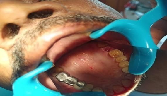 |
Fig 1: pre-operative intraoral photograph showing small oroantral fistula
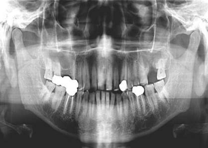
Fig 2: pre-operative orthopantomogram
The oroantral fistula was decided to be closed using a palatal pedicle flap surgically under local anesthesia. Buccal and palatal anesthesia was administered using 2% lignocaine with 1:200,000 adrenaline.
The fistulous tract was debrided, and the surrounding bone was freshened. A crevicular incision was placed on the palate, extending to the first premolar area, using a No. 15 BP blade (Fig. 3). A palatal connective tissue pedicle flap was prepared by careful dissection of the palatal mucosa and rotated gently to achieve tension- free closure of the oroantral communication (Fig. 4). The superficial epithelial layer was repositioned to its normal position (Fig. 5). Postoperatively, the patient was prescribed antibiotics and analgesics for 5 days.
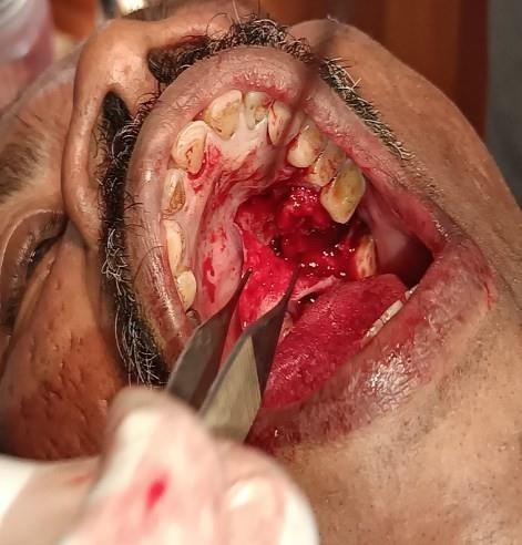 |
Fig 3: Preparation of the palatal pedicle flap
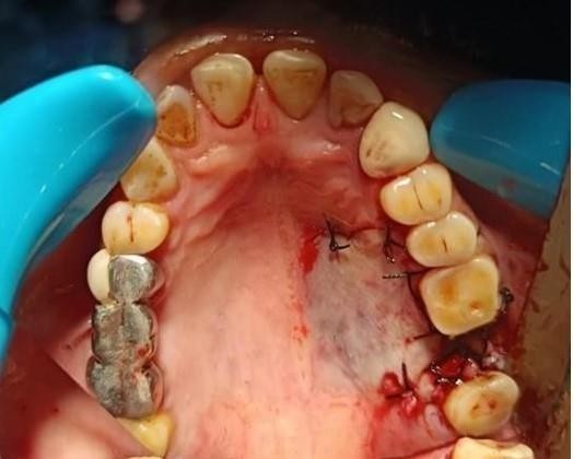 |
Fig 4: The flap sutured hermetically to the surgical site
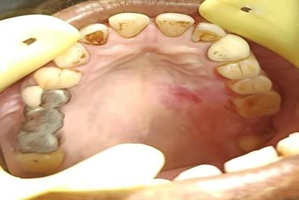
Figure 5: Surgical site after 4weeks showing good healing response
DISCUSSION
OAF is usually asymptomatic; however, some cases may present with epistaxis, passage of fluid or air, pain in and around the involved sinus area, alteration of voice, wheezing during speech, free drainage of fluids through the oral fistula, mucopurulent nasal discharge, antral polyp, altered taste, halitosis, ear ache, etc. OAF can be clinically detected by observing fogging of a mouth mirror when placed at the opening. It is further confirmed using the Valsalva test, in which the patient gently expels air against closed nostrils while keeping the mouth open; the passage of air or blood at the postoperative site is confirmatory of OAF. However, a negative test does not necessarily rule out antral perforation, and small OAFs may not always be easily detected. Radiographs and CT scans confirm the presence of OAF while also determining its size, location, and degree of sinus involvement.[6] Large non healed OAF is treated by surgical techniques using flaps from either buccal or palatal tissues, using buccal fat pad, tongue flaps, bone grafts and alloplastic material like hydroxyapatite, soft polymathic methacrylate, resorb able collagen membranes, gold foil and gold plates. The choice of technique depends on the size, location, and seniority of the OAF, patient age, and medical comorbidities and upon surgeon’s experience and technical skills.[4] Vestibular sliding flap described by Rahman 1936 is the oldest and the most frequently used technique for closure of OAF. The success rate is very high due to the wide base of the flap giving adequate vascularization. Buccal fat pad (BFP) flap requires minimal dissection to harvest, mobilize flap and has high success rate attributed to rich vascularization from maxillary artery.[7] The palatal connective tissue pedicle flap proved to be an effective technique for closing the oroantral fistula in this case. The flap's excellent vascularity and flexibility allowed for tension-free closure, promoting uneventful healing. The minimal donor site morbidity and absence of significant postoperative complications further support the use of this technique. This contrasts with the belief that palatal flaps cause great morbidity due to open area it creates in the palate which entails longer post-operative care along with significant discomfort to the patient.[8] As a musician who played a wind instrument, the patient's occupation necessitated a reliable closure to prevent air leakage and ensure optimal performance the success of this procedure highlights the potential benefits of using palatal connective tissue pedicle flaps in similar cases, particularly where occupational demands require precise functional restoration. Several surgical techniques for OAF closure have been introduced in the literature. Buccal and palatal flaps are commonly used methods, while the other local flaps are mostly variations of the two techniques. The size and location of the defect, the presence of acute or chronic infection in the sinus, and the absence of sufficient vestibular depth or keratinized tissue surrounding the defect are all determinately important factors for the preference of surgical technique to close the defect. Additionally, during planning of the flap design, the surgeon should consider whether it is immediate or delayed, whether there is thick and healthy tissue surrounding the defect, and whether the patient is healthy or medically compromised.[9] Full-thickness rotational palatal flaps offer benefits like keratinized tissue, vestibular depth preservation, and improved healing. However, they can limit rotation and cause kinking, compromise vascular supply and cause venous congestion. Kruger recommends excising a V- shaped section to prevent folding. The technique exposes the hard palate's bony structure, causing pain, burning, and edema. Necrosis risk is high, especially in compromised patients. Post-operatively, a palatal stent is recommended to stabilize the flap.[10] Dargin et al. developed a modified submucosal connective tissue flap for OAF repair, allowing better manipulation and adaptation in the closure of donor sites. This technique eliminates folding or dog-ear formation and requires no palatal acrylic plate postoperatively.[11] Ito and Hara modified the pedicle palatal flap, dividing it into an upper epithelial layer and underlying connective tissue layer. Healing occurs within 1 month, but has disadvantages like difficulty in dissection, blood supply injury, and surgeon experience.[12]
CONCLUSIONThe palatal connective tissue pedicle flap technique is a valuable approach for managing OAFs, offering a high success rate and minimal complications. This case report highlights the effectiveness of this technique in achieving successful closure of OAFs.
References
REFERENCES: